Health
How Do you Test for Blocked Arteries in Your Neck?

Testing for blocked arteries in the neck, commonly known as carotid artery disease, is critical for determining cardiovascular health and diagnosing potential stroke or other issues. Several diagnostic methods are available to assess the status of the carotid arteries and the extent of obstruction.
In this article, we will look at the numerous ways for testing for blocked arteries in the neck, including non-invasive imaging techniques like carotid ultrasonography, computed tomography angiography (CTA), magnetic resonance angiography (MRA), and digital subtraction angiography.
Understanding these testing procedures is critical for healthcare professionals and individuals who are at risk of or have symptoms of blocked arteries in the neck, as early detection and treatment can help prevent major problems and improve overall cardiovascular health.

Importance of early detection and diagnosis for Blocked Arteries in Your Neck
Early detection and diagnosis of blocked arteries in the neck, known as carotid artery disease, is crucial in preventing major complications including stroke and transient ischemic episodes (TIAs), often known as mini-strokes. Identifying the prevalence and severity of carotid artery blockages enables healthcare practitioners to intervene quickly and adopt appropriate treatment measures to reduce the risk of stroke and its debilitating effects.
A timely diagnosis allows healthcare practitioners to implement preventative treatments and lifestyle changes aimed at slowing disease progression. Furthermore, early detection enables the implementation of focused medical interventions, such as medication management, to reduce risk factors such as high blood pressure, high cholesterol, and diabetes.
Furthermore, early diagnosis allows for the consideration of interventional techniques or surgical interventions when necessary, such as carotid endarterectomy or carotid artery stenting, to remove or bypass the blockage and restore normal blood flow to the brain. These therapies are most successful when implemented before to the onset of a stroke or other neurological problems.
Individuals with risk factors such as hypertension, diabetes, high cholesterol, smoking, obesity, or a family history of cardiovascular disease should undergo regular carotid artery disease screening. Routine screenings and diagnostic testing can detect artery blockages early on, allowing for timely intervention and proactive management to avoid negative results.
To summarize, early detection and diagnosis of blocked arteries in the neck is critical for implementing preventative measures, commencing suitable therapies, and lowering the risk of stroke and its sequelae. Individuals can take proactive actions to protect their cardiovascular health and overall well-being by scheduling frequent exams and seeking medical assistance as soon as any troubling symptoms appear.

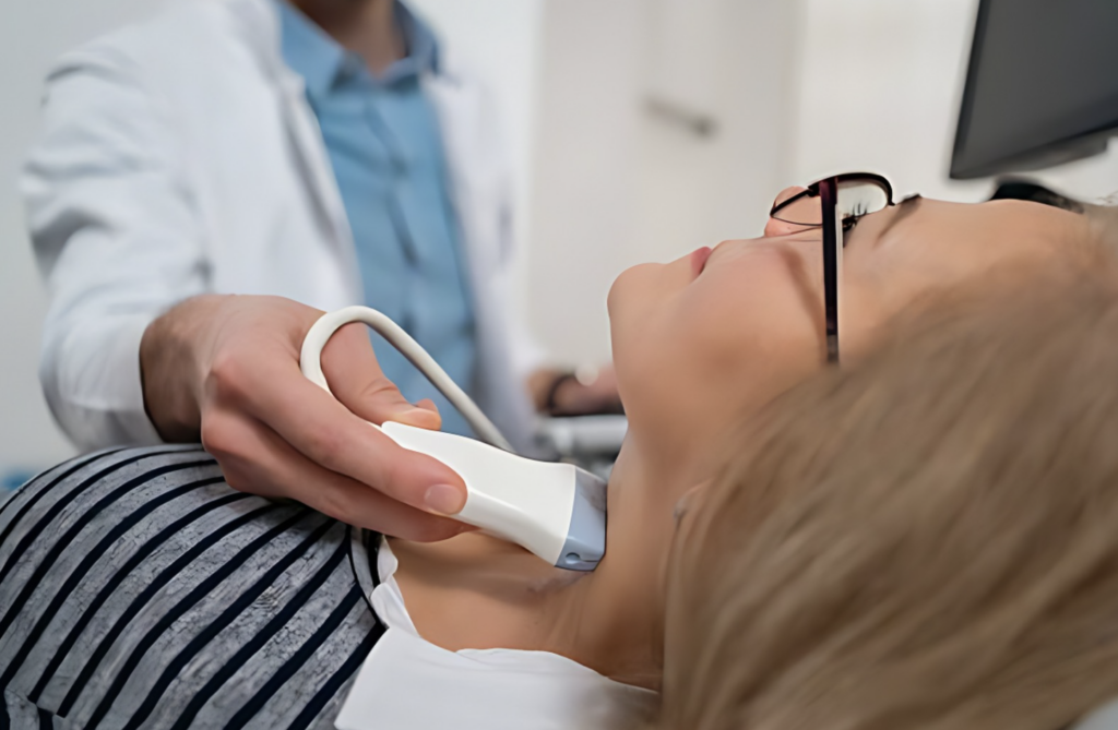
Carotid ultrasound: A non-invasive and painless test
Carotid ultrasound is a non-invasive imaging technique used to diagnose carotid artery disease, which is defined by narrowing or blockage of the carotid arteries that carry blood to the brain. A carotid ultrasound uses high-frequency sound waves to create detailed pictures of the carotid arteries and monitor blood flow.
This diagnostic process enables healthcare providers to determine the presence and degree of blockages, as well as any plaques or other abnormalities in the carotid arteries. Carotid ultrasonography assesses the risk of stroke and determines the best treatment option by evaluating blood flow velocity and detecting areas of narrowing or stenosis.
Carotid ultrasound is a painless procedure that is usually performed in a clinical setting by a qualified technician or healthcare provider. It entails putting gel to the neck’s skin and using a handheld equipment known as a transducer to collect images of the carotid arteries from various angles.
A carotid ultrasound can help healthcare providers make informed decisions about treatment options, such as lifestyle changes, medication management, or surgical interventions, to reduce the risk of stroke and other complications caused by carotid artery disease.
Overall, carotid ultrasound is an important tool in the diagnosis and management of carotid artery disease, enabling for early detection, risk stratification, and focused intervention to maintain cardiovascular health and avoid bad events such as stroke.

CT angiography: Detailed imaging of the blood vessels
CT angiography (CTA) is a diagnostic imaging procedure that examines the carotid arteries for anomalies or blockages that may suggest carotid artery disease. This non-invasive method uses computed tomography (CT) scanning and a contrast dye injection to provide a detailed view of the blood arteries.
In a CT angiography procedure for carotid artery disease, a contrast dye is injected into a vein, generally in the arm, to highlight the blood vessels on the CT pictures. The patient is then placed on a CT scanner table, which passes through a doughnut-shaped machine that captures cross-sectional images of the neck.
The CT scanner produces comprehensive images of the carotid arteries, allowing doctors to determine the presence and severity of blockages, narrowing, or plaque formation. These images show the flow of blood via the carotid arteries, which aids in the diagnosis of carotid artery disease and the development of an effective treatment strategy.
CT angiography is especially effective for analyzing the morphology of the carotid arteries, determining the location and degree of blockages, and calculating the risk of stroke. It is frequently used in patients who have symptoms of carotid artery disease, such as transient ischemic episodes (TIAs) or stroke, as well as those who are at high risk of cardiovascular events.
Overall, CT angiography is an effective method for diagnosing and managing carotid artery disease, providing healthcare clinicians with critical information to guide treatment decisions and lower the risk of stroke and other problems.

Head CT scan: Detecting blockages and other abnormalities
A head CT scan, while not intended to diagnose carotid artery disease, might indirectly reveal information on the status of the carotid arteries. A head CT scan uses X-rays to take images of the brain and surrounding tissues in order to detect anomalies such as tumors, hemorrhage, or signs of a stroke.
While a head CT scan can not directly visualize the carotid arteries, it can occasionally reveal secondary symptoms of carotid artery disease, such as evidence of a stroke or transient ischemic attack (TIA) caused by decreased blood supply to the brain due to carotid artery blockage or constriction. A head CT scan may also reveal calcifications in the carotid arteries, indicating the presence of atherosclerosis, the most prevalent cause of carotid artery disease.
However, for a more complete assessment of the carotid arteries, specialist imaging techniques such as carotid ultrasound or CT angiography (CTA) are usually chosen. These imaging techniques are specifically intended to analyze the structure and blood flow of the carotid arteries, providing more precise information regarding the amount of blockages or narrowing.
In summary, while a head CT scan can provide some information about the presence of carotid artery disease, it is not the major imaging modality utilized to diagnose or evaluate this illness. Instead, carotid ultrasonography or CT angiography are the preferable procedures for directly viewing and measuring the carotid artery.

Magnetic Resonance Angiography (MRA): Visualizing blood flow
Magnetic Resonance Angiography (MRA) is a non-invasive imaging technology that combines magnetic fields and radio waves to provide detailed views of blood vessels, including the carotid arteries. MRA is often used to assess carotid artery disease by producing high-resolution pictures of the arteries and detecting any blockages, narrowing, or irregular blood flow.
During an MRA procedure for carotid artery disease, a contrast agent may be injected into the circulation to improve blood vessel visibility. This allows radiologists to detect and analyze the severity of any stenosis (narrowing) or plaque buildup in the carotid arteries, both of which are signs of carotid artery disease.
MRA has several advantages for evaluating carotid artery disease, including its non-invasive nature, ability to produce detailed images without exposing patients to ionizing radiation, and absence of the potential allergic reactions associated with contrast agents used in other imaging modalities such as CT angiography.
Overall, MRA is an effective diagnostic tool for assessing carotid artery disease, providing doctors with critical information for guiding treatment decisions and treatments targeted at lowering the risk of stroke and other cardiovascular problems associated with this illness.

MRI scan: Evaluating the health of blood vessels
Magnetic Resonance Imaging (MRI) is a non-invasive imaging technique that creates detailed images of the body’s interior systems, including the carotid arteries, using strong magnets and radio waves. An MRI scan can be used in the context of carotid artery disease to check the health and integrity of the arteries, as well as to detect any anomalies such as plaque buildup or constriction (stenosis) that may obstruct blood flow.
An MRI scan for carotid artery disease involves the patient lying on a table that slides into the MRI scanner, which surrounds them with a strong magnetic field. Radio waves are then directed to the body, causing hydrogen atoms in its tissues to generate signals that the MRI equipment detects and converts into detailed images.
An MRI scan of the carotid arteries can reveal important information regarding the location and severity of any blockages or anomalies, allowing healthcare providers to properly diagnose and manage carotid artery disease. Furthermore, MRI scans are non-invasive and do not use ionizing radiation, making them a safe option for imaging the carotid arteries.
Overall, MRI scans are a useful tool in the diagnosis of carotid artery disease, providing doctors with critical information to guide treatment decisions and treatments targeted at lowering the risk of stroke and other consequences.

Cerebral angiography: Invasive but highly accurate
Cerebral angiography, also known as carotid angiography or carotid arteriography, is a diagnostic imaging technique that visualizes the carotid arteries and measures blood flow in the brain. It is often used to assess carotid artery disease, such as stenosis (narrowing) or blockages in the carotid arteries, which can raise the risk of stroke.
During cerebral angiography, a contrast dye is injected into the carotid arteries via a catheter put into a blood vessel in the groin or arm. The contrast dye reveals blood veins in the brain, allowing radiologists to produce detailed X-ray images known as angiograms.
These angiograms reveal detailed information about the anatomy and status of the carotid arteries, such as the existence and amount of plaque buildup, narrowing, or blockages. This information is critical for detecting carotid artery disease and determining the best treatment option.
Cerebral angiography is regarded as the gold standard for diagnosing carotid artery disease because it produces high-resolution pictures and enables for direct imaging of the blood vessels. However, it is an invasive technique with potential hazards, such as bleeding, allergic responses to the contrast dye, and stroke, which are normally modest when conducted by skilled healthcare professionals.
Overall, cerebral angiography is critical in the diagnosis and management of carotid artery disease, giving significant information to guide treatment decisions and treatments targeted at lowering the risk of stroke and other problems associated with the condition.

Choosing the right test for your condition
Choosing the right test for carotid artery disease depends on various factors, including your symptoms, medical history, and the severity of your condition. Here are some considerations to help you and your healthcare provider decide which test is most appropriate:
- Symptoms: If you’re having symptoms of carotid artery disease, such as transient ischemic episodes (TIAs) or stroke-like symptoms, your doctor may recommend more immediate testing to determine the extent of blockage in your carotid arteries.
- Risk Factors: Age, smoking, high blood pressure, high cholesterol, diabetes, and a family history of vascular disease all raise your chances of getting carotid artery disease. If you have these risk factors but no symptoms, your doctor may suggest screening tests to determine your risk.
- Diagnostic Accuracy: Each carotid artery disease test has a certain amount of accuracy in finding blockages and narrowing of the carotid arteries. When deciding which test is best for you, your healthcare professional will assess its sensitivity and specificity.
- Invasiveness: Certain tests, such as cerebral angiography, are more intrusive and have a higher risk of problems than non-invasive diagnostics, such as carotid ultrasonography. Your healthcare professional will assess the risks and benefits of each test based on your general health and medical history.
- Availability and Accessibility: The availability of specific tests may differ depending on your region, healthcare facility, and insurance coverage. When recommending a specific test, your healthcare professional will take into account these practical aspects.
- Patient Preference: Your preferences and degree of comfort with various testing techniques may also factor into the selection. Make sure to share any concerns or preferences you have with your healthcare professional.
Ultimately, the choice of test for carotid artery disease will depend on a comprehensive evaluation of your individual circumstances. Your healthcare provider will work with you to determine the most appropriate diagnostic approach to effectively assess and manage your condition.
Disclaimer: Please note that Discoverybody has taken great care to ensure that all information provided is comprehensive and up-to-date. However, you should not use this article as a substitute for the expertise that a licensed healthcare professional can offer. It’s always a good idea to talk to your doctor before taking any medication.
Sources Expanded:
Phifer, A. (2024, February 27). Your guide to early detection and prevention of carotid artery disease – Baylor College of Medicine Blog Network. Baylor College of Medicine Blog Network. https://blogs.bcm.edu/2024/02/28/your-guide-to-early-detection-and-prevention-of-carotid-artery-disease/
Carotid ultrasound – Mayo Clinic. (2023, January 12). https://www.mayoclinic.org/tests-procedures/carotid-ultrasound/about/pac-20393399#:~:text=A%20carotid%20ultrasound%20is%20done,that%20circulate%20in%20the%20bloodstream.
Professional, C. C. M. (n.d.). Carotid Angiography. Cleveland Clinic. https://my.clevelandclinic.org/health/diagnostics/16842-carotid-angiography
Diagnosis. (n.d.). SCAI – Seconds Count. https://www.secondscount.org/condition/carotid-artery-disease/diagnosis#:~:text=A%20CT%20scan%20can%20give,to%20see%20the%20blood%20vessels.
Magnetic resonance angiography (MRA). (n.d.). Stanford Health Care. https://stanfordhealthcare.org/medical-conditions/blood-heart-circulation/carotid-artery-disease/diagnosis/magnetic-resonance-angiography.html
Carotid Artery Disease Imaging. (2022, August 12). Yale Medicine. https://www.yalemedicine.org/conditions/carotid-artery-disease-imaging#:~:text=Usually%2C%20it%20provides%20a%20clear,(CT)%20are%20also%20needed.
Professional, C. C. M. (n.d.). Cerebral Angiogram. Cleveland Clinic. https://my.clevelandclinic.org/health/diagnostics/13476-cerebral-angiogram
Cerebral Angiography. (n.d.). Radiologyinfo.org. https://www.radiologyinfo.org/en/info/angiocerebral
Trusted Health, Wellness, and Medical advice for your well-being